
Prevalence
Venous leg ulcers (VLUs) are one of the more common types of leg ulcers, accounting for up to 70% of all chronic leg ulcers.1 Prevalence increases by 4% for people over the age of 65.2 VLUs typically appear in the gaiter area of the leg, above the malleolus to one inch below the knee. VLUs are shallow but large wounds with irregular borders.

Risk Factors
The primary risk factors for VLUs include age, obesity, deep venous thrombosis, family history of venous disease, advanced age, lower extremity and orthopedic procedures.3 African Americans, Asians, and non-Hispanic White are at high risk.4

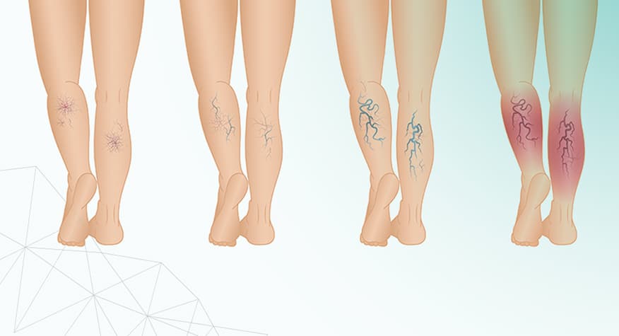
Pathophysiology
VLUs develop when the valves inside the veins of the lower extremities do not work sufficiently, resulting in backflow. This process leads to blood pooling in the veins (venous hypertension) followed by edema and eventually the development of an ulcer.5

Assessment
The clinical diagnosis of VLUs is based on physical examination. Ulcers in which the etiology is unclear, venous duplex can assist in making the diagnosis; a biopsy may also be considered.

Prevention and Treatment
Patient education, weight reduction, smoking cessation, diet modification and supplementation are recommended for prevention of leg ulcers. Multilayer compression therapy is the gold standard for VLUs. Prior to the application of any compression wrap, vascular screening should be performed. Standard compression wraps require an ABI > 0.8.6,7 Light wraps are available for use in patients with ABI as low as 0.6.8 Change compression wraps should be changed at least weekly.
References
- Pugliese DJ. Infection in Venous Leg Ulcers: Considerations for Optimal Management in the Elderly. Drugs Aging. 2016;33(2):87-96. doi:10.1007/s40266-016-0343-8.
- Vasudevan B. Venous leg ulcers: Pathophysiology and Classification. Indian Dermatol Online J. 2014;5(3):366-370. doi:10.4103/2229-5178.137819.
- Abbade LP, Lastória S, Rollo Hde A. Venous ulcer: clinical characteristics and risk factors. Int J Dermatol. 2011;50(4):405-411. doi:10.1111/j.1365-4632.2010.04654.x.
- Michael H. Criqui, Maritess Jamosmos, Arnost Fronek, Julie O. Denenberg, Robert D. Langer, John Bergan, Beatrice A. Golomb, Chronic Venous Disease in an Ethnically Diverse Population: The San Diego Population Study, American Journal of Epidemiology, Volume 158, Issue 5, 1 September 2003, Pages 448–456, https://doi.org/10.1093/aje/kwg166.
- Chi YW and Raffetto JD. Venous leg ulceration pathophysiology and evidence based treatment. Vascular Medicine. 2015; 20(2):168-181.
- Harding K, Dowsett C, Fias L, et al. Simplifying venous leg ulcer management. Consensus recommendations. Wounds Int. 2015;6(2):54.
- Ratliff CR, Yates S, McNichol L, Gray M. Compression for primary prevention, treatment, and prevention of recurrence of venous leg ulcers: an evidence-and consensus-based algorithm for care across the continuum. J Wound Ostomy Continence Nurs. 2016;43(4):347-64.
- Bernatchez SF, Peterson L Fife CE. Compression therapy: the key to unlocking VLU healing. Today’s Wound Clinic. 2017; 11(12).